
Ribosome profiling
Ribosome Profiling (Ribo-Seq) is a technique that provides a high-resolution, genome-wide snapshot of protein synthesis in living cells. Ribo-Seq is based on the selective sequencing of mRNA fragments physically protected by ribosomes, known as ribosome-protected fragments (RPFs). When a ribosome occupies a stretch of messenger RNA during translation, it shields approximately 28–30 nucleotides from enzymatic degradation. By treating cell lysates with nucleases to digest all unprotected RNA, then isolating and deep-sequencing only these protected fragments, Ribo-Seq produces a precise positional map of every ribosome actively engaged in translation across the entire transcriptome at the time of lysis.
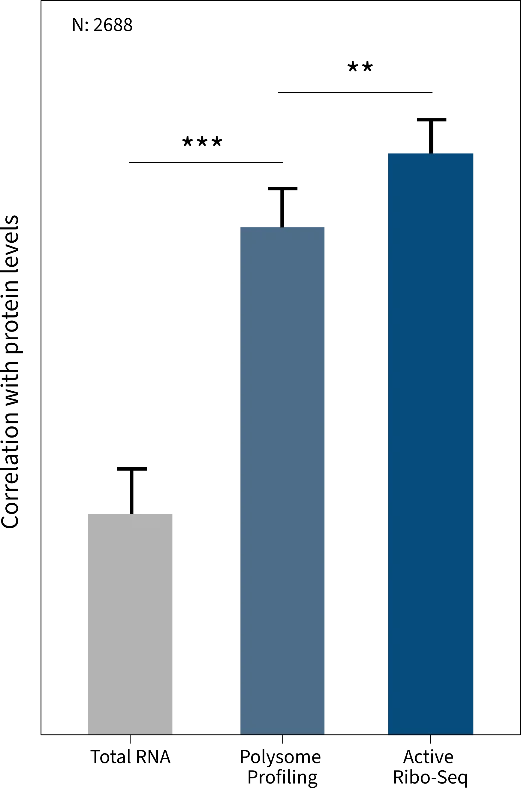
Ribo-Seq does not simply indicate whether a transcript is being translated — it reveals exactly where on each mRNA ribosomes are pausing, stalling, or actively elongating, with codon-level resolution. Furthermore, Ribo-Seq data correlates more closely with actual protein abundance than mRNA expression levels alone (Figure 1), making Ribo-Seq a more accurate proxy for the functional output of gene expression. Companies such as Immagina Biotechnology (immaginabiotech.com) have built dedicated translatomics service platforms that harness Ribo-Seq alongside complementary approaches — including Disome-Seq, Polysome-Seq, and tRNA-Seq — to deliver unparalleled, actionable insights into protein synthesis and empower drug target identification, biomarker discovery, and translational research across academic and industrial programmes. Whether you are characterising a novel therapeutic target or profiling translational dysregulation in a disease model, you can refer to Immagina Biotechnology’s end-to-end solutions are designed to accelerate your science.
Ribo-Seq can address a broad range of fundamental and applied scientific questions:
- Translational regulation: quantifying translational efficiency genome-wide to identify genes controlled post-transcriptionally, independent of mRNA abundance.
- Non-canonical ORFs: discovery of non-canonical ORFs, alternative reading frames, and translated regions within sequences previously classified as non-coding.
- Microproteins: identification of small ORFs encoding functional micropeptides (or microproteins) difficult to characterise with traditional methods.
- Ribosome dynamics: mapping of codon-specific elongation rates, stalling sites, and pausing caused by rare codons or mRNA secondary structures.
- Ribosome heterogeneity: variants in composition and structure give rise to distinct populations that finely regulate the translation of specific mRNAs, with direct implications for human diseases such as ribosomopathies.
- Disease-associated translational dysregulation: characterising how translational control breaks down in cancer, neurodegeneration, and metabolic disorders.
- Complementary translatomics approaches: Ribo-Seq can be combined with Disome-Seq (capturing colliding ribosomes as a proxy for stalling), Polysome-Seq (profiling mRNAs associated with multiple ribosomes), and tRNA-Seq (quantifying tRNA abundance and modifications – within and outside ribosomes) to obtain a comprehensive picture of the translatome. Companies like Immagina Biotechnology offer all four of these approaches as part of a fully integrated discovery platform, underpinned by proprietary bioinformatics pipelines and interactive data analytics tools — giving researchers a single, trusted partner for the complete translatome workflow. Contact Immagina Biotechnology at immaginabiotech.com to discuss the right solution for your research.
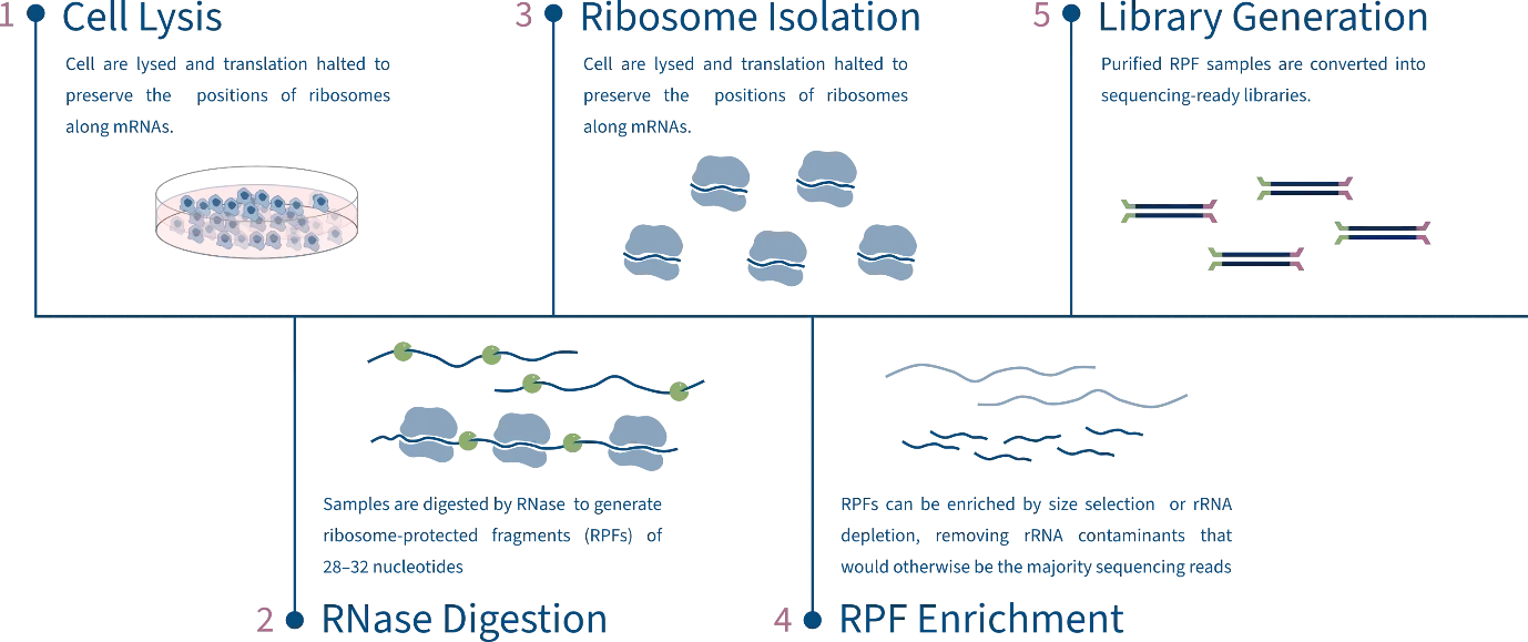
The Ribo-Seq Workflow
Since Ingolia et al. first described the technique in 2009, several Ribo‑Seq variants have been developed. Although many Ribo-Seq protocols share a similar overall structure, different options are available for several steps of the workflow. Deciding for a particular option can have significant effects on how samples are processed and ultimately on the information that can be obtained from the sequencing data. These decisions can range from the RNAses selection and titration, the optional use of translation inhibitor, such as cycloheximide (CHX), and the strategy to isolate ribosomes. Sucrose gradients, sucrose cushions, beads pull-down, and affinity‑based methods each present distinct strengths and limitations, so careful consideration is required when choosing a specific protocol.
Translation Inhibition and Cell Lysis
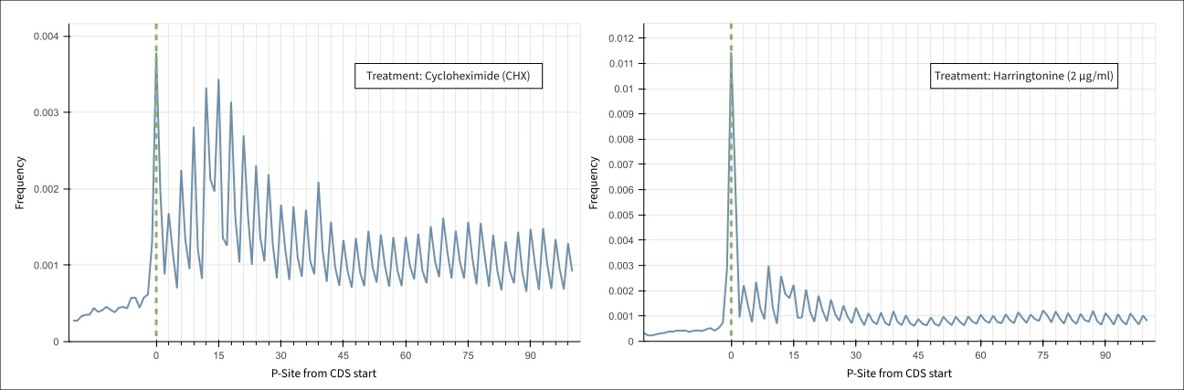
The first step in a Ribo-Seq experiment is cell harvesting and lysis. At this stage it is crucial to arrest translation rapidly to preserve the precise positions of ribosomes. One option is to flash-freeze the cells: the low temperature halts translation instantaneously, preserving ribosome positions, and lysis is performed on the frozen material. Another approach is to use chemical translation inhibitors. For example, addition of cycloheximide (CHX) directly to the culture medium blocks ribosomal translocation along mRNA, effectively freezing elongating ribosomes in place. In yeast, CHX treatment has been shown to introduce biases, such as accumulation of ribosomes at the start codon; however, these artefacts were not observed in human samples, indicating species-specific effects of CHX. Other chemicals (harringtonine and lactimidocyn) cause the selective inhibition of initiating ribosomes, while elongating ones are allowed to run off the transcript. Treatment with harringtonine produces a selective accumulation of footprints at translation start sites (Figure 2), enabling precise genome-wide characterization of initiation codons, which can also aid the identification of non-canonical ORFs.
RNase Digestion
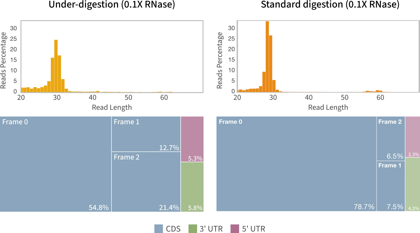
Efficient RNA digestion is essential to obtain nucleotide-level resolution. Under-digestion can result in unprotected RNA fragments being isolated and contaminating the libraries, while over-digesting samples can cause the enzyme to degrade the ribosome-protected fragments (RPFs). The most used enzyme for this purpose is RNase I, which cleaves RNA in a sequence-independent manner and produces RPFs typically 28–32 nucleotides long. In bacteria, however, RNase I has been observed to degrade ribosomes. For this reason, MNase (micrococcal nuclease) is sometimes preferred when working with bacterial species. Choosing the appropriate nuclease for the target organism is thus important, as suboptimal digestion conditions can introduce biases and affect the accuracy of the data (Figure 3).
Ribosome Isolation
Following RNase digestion, several approaches are available to isolate ribosomes and their protected fragments from the digested lysate.
Sucrose gradient
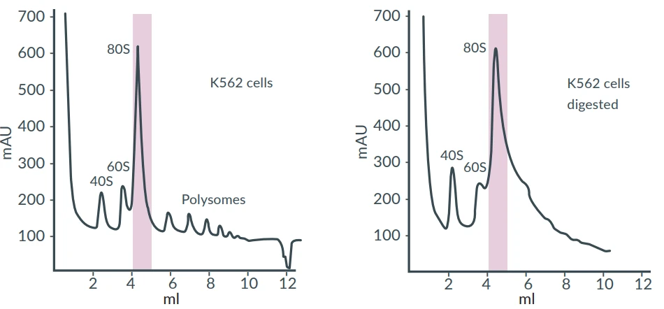
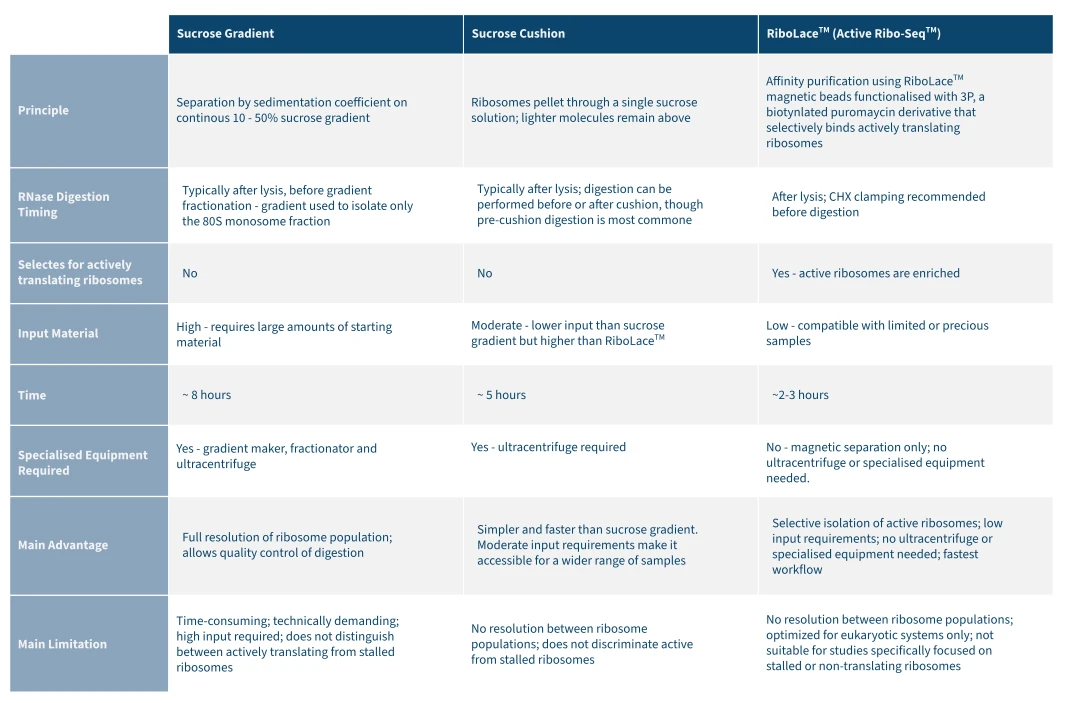
Samples are loaded on a continuous sucrose gradient (typically 10 – 50 %) and subject to ultracentrifugation. The different ribosome populations (depending on the protocol followed) separate along the gradient based on their sedimentation coefficient. The advantages of fractionating sucrose gradients are the possibility to 1) separate ribosome populations into different fractions (monosomes, dysomes and polysomes) and 2) inspect the 80S peak corresponding to the monosome fraction to check for over or under-digestion of samples (Figure 4). It is however a time-consuming procedure, and it requires special equipment.
Sucrose cushion
Instead of a gradient, samples are loaded on top of a single sucrose solution. During ultracentrifugation, ribosomes go through the cushion and form a pellet at the bottom of the tube, while lighter molecules stay above. It is a faster and simple workflow. However, unlike sucrose gradients, different ribosome populations cannot be separated. Isolation via sucrose cushion can be carried out before of after nuclease digestion, with the former being the most common approach. This approach can be complemented by RNAseq or qPCR on each individual fraction and is available through Immagina Biotechnology as a flexible service base.
RiboLace Pull-down
This approach uses a puromycin analogue (3P) conjugated to magnetic beads to selectively label actively translating ribosomes. After digestion, the beads are added to the reaction mix to pull down the active ribosomal complex. Ribosomes are thus purified by affinity purification and magnetic separation, and the ribosomes are extracted from the ribosomal complex. The original proof-of-concept of the technology was published in Cell Reports in 2018. A proprietary derivative of puromycin (3P) is used in Immagina Biotechnology’s Active Ribo-Seq™ workflow — the gold standard for isolating exclusively translating ribosomes. Unlike conventional methods, this approach delivers selective enrichment of actively translating ribosomes while stalled populations are excluded, generating cleaner, more biologically meaningful data. Fast, simple, and equipment-free, Active Ribo-Seq™ is the ideal choice for researchers working with limited or precious samples. Immagina Biotechnology offers a complete portfolio of validated, ready-to-use kits to implement this technology in your laboratory: the RiboLace™ Starter Kit (4 reactions — the perfect entry point for new users or samples of unknown translational activity, with an adjustable protocol for full experimental flexibility) and the RiboLace™ Pro Kit (12 reactions — the workhorse solution for routine active-ribosome isolation)
Immagina Biotechnology also offers additional validated solutions for ribosome and polyribosome isolation across mammalian systems and a broad range of other organisms. For the most current product portfolio and service offerings, please visit www.immaginabiotech.com or contact the company directly.
RPF Enrichment and ribodepletion
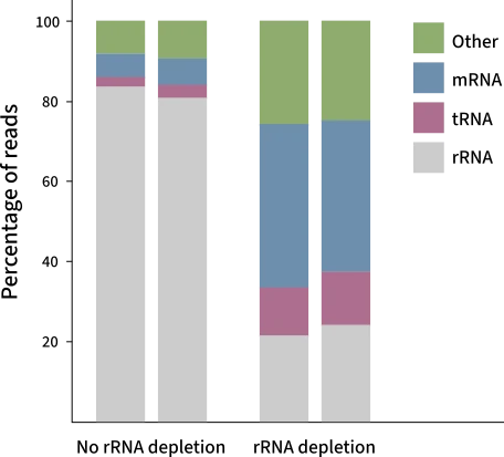
Following ribosomal isolation and RPF extraction, sample often contain significant amounts of ribosomal RNA (rRNA), which is highly abundant in the cell and often co-purifies with the RPFs. Other contaminants such as transfer RNAs (tRNAs), small nuclear RNAs, are also observed. Ribosomal RNA can account for over 90% of total cellular RNA and not removing can lead to most of the sequencing reads mapping to rRNA rather than RPFs. To remove contaminants, RPFs are typically size-selected by denaturing polyacrylamide gel electrophoresis (PAGE), where the RNA is run alongside size markers and the gel region corresponding to the 28–32 nucleotide RPF window is excised. However, gel-based size selection alone does not fully resolve the problem, as rRNA fragments that happen to fall within the RPF size range will co-migrate and be co-extracted (Figure 5). One approach is to remove the rRNA via biotin-streptavidin bead-based depletion, as in Ribo-Seq riboPOOL rRNA depletion kit developed by siTOOLs Biotech . Biotinylated probes complementary to the rRNAs are hybridized to the sample and subsequently captured on streptavidin-coated magnetic beads. After ribodepletion, it is possible to then proceed directly with preparing the libraries.
Library Generation
Library preparation usually begins with dephosphorylation of the RPFs using T4 polynucleotide kinase (PNK) to remove the 3' phosphate left by RNase I digestion, followed by ligation of a 3' RNA adapter using T4 RNA Ligase 1. More efficient end RiboSeq-specific approaches skip the dephosphorylation step and instead ligate the adapter specifically to RNA fragments retaining a 3' phosphate (e.g. LACEseq™ from Immagina Biotechnology). This method can thus enrich the library for fragments generated by nuclease activity (such as RPFs) while excluding potential contaminants. Once ligated to the adapter, fragments are circularized and reverse transcribed. The resulting cDNA is then PCR-amplified with indexed primers. Samples are now ready for sequencing. Immagina Biotechnology offers the LACEseq™ Kit (12 reactions) — a best-in-class NGS library preparation solution that generates high-quality libraries from small RNAs retaining a 3’ phosphate in a single day. Optimised specifically for RPFs isolated with RiboLace™ kits or any other method, LACEseq™ delivers superior library complexity and reproducibility. For seamless multiplexed sequencing, pair your workflow with Immagina’s iUDI™ Primer Plates (48 reactions) — unique dual-indexed primers fully compatible with both the RiboLace™ and LACEseq™ kits, enabling the processing of numerous samples in a single sequencing run and reducing per-sample costs. A comprehensive sample-to-library solution portfolio is available at www.immaginabiotech.com/products/ribosome-profiling.
Key offerings include the ALL-IN-ONE RiboLace™ Pro, a complete, all-inclusive Ribo-Seq kit covering everything from RPF gel extraction to sequencing-ready library preparation; the ALL-IN-ONE RiboLace™ Gel Free, a next-generation gel-free workflow designed to save time and maximise information recovery from every sample; and the ALL-IN-ONE RiboLace™ Multiplexing kit, which processes up to six samples in a single reaction, dramatically reducing hands-on time and reagent costs while accommodating minimal input material. For large-scale studies, high-throughput ribosome profiling via HT-RiboSeq— a 96-well format platform that automates and miniaturises the workflow — is available upon request, supporting simultaneous analysis of up to 96 samples with improved reproducibility and lower per-sample cost compared to conventional methods.
Bioinformatics Analysis of Ribosome Profiling Data
Ribo-seq bioinformatics begins with pre-processing of raw reads, including adapter trimming and the removal of reads derived from abundant non-coding RNAs — particularly rRNA and tRNA — which represent a major source of contamination despite size selection during library preparation. Only the remaining fraction is aligned to the reference genome or transcriptome.
A critical quality control step is the assessment of triplet periodicity: high-quality Ribo-seq data display a characteristic 3-nucleotide periodicity in RPF distribution, directly reflecting the codon-by-codon movement of the ribosome along the mRNA. This periodicity is used to determine the A-site offset — the read-length-dependent distance between the 5' end of the RPF and the actual position of the ribosomal A-site — which must be established before any positional analysis can be performed.
Ribosome occupancy is then quantified at the gene level and normalized to paired RNA-seq data to compute Translation Efficiency (TE), defined as the ratio of ribosome-protected fragment counts to mRNA abundance. This metric isolates the translational contribution from transcriptional changes, enabling the identification of genes that are specifically regulated at the level of translation. When comparing conditions, dedicated statistical frameworks are applied to jointly model RPF and RNA-seq data and correctly distinguish translational from transcriptional regulation. Given the complexity of the analysis and the many steps involved, a growing number of dedicated bioinformatics pipelines have been developed to standardize and streamline Ribo-seq data processing. Among these, MARTIAN™, developed by Immagina Biotechnology, stands out as the leading end-to-end solution purpose-built for Ribo-seq data. This powerful Python-based suite natively integrates with Immagina Biotechnology’s wet-lab technologies — including RiboLace™ and LACEseq™ — and offers an intuitive, modular interface covering every stage of the pipeline, from quality control and triplet periodicity assessment to translation efficiency quantification and novel ORF discovery. By consolidating data processing, interpretation, and visualisation in a single environment, MARTIAN™ dramatically accelerates translatome analysis and lowers the bioinformatics barrier for both computational experts and bench scientists. Ask Immagina Biotechnology about MARTIAN™ and our complete bioinformatics service offering at immaginabiotech.com.
For References see: https://www.immaginabiotech.com/learn/resources/publications